Vui lòng dùng định danh này để trích dẫn hoặc liên kết đến tài liệu này:
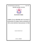
http://dulieuso.hmu.edu.vn/handle/hmu/1835| Nhan đề: | Nghiên cứu đặc điểm hình ảnh và giá trị của cộng hưởng từ 1,5 Tesla trong chẩn đoán và tiên lượng nhồi máu não giai đoạn cấp tính. |
| Tác giả: | Nguyễn Duy, Trinh |
| Người hướng dẫn: | GS.TS. Phạm Minh, Thông GS.TS Lê Văn, Thính |
| Từ khoá: | 62720311;Chẩn đoán hình ảnh |
| Tóm tắt: | THÔNG TIN TÓM TẮT NHỮNG KẾT LUẬN MỚI CỦA . LUẬN ÁN TIẾN SĨ. . Những kết luận mới của luận án:. 1 Đặc điểm hình ảnh cộng hưởng từ nhồi máu não cấp tính:. - Thể tích nhồi máu não trung bình nhóm nghiên cứu là 45,6 ± 67,4 cm3. Thể tích nhồi máu trung bình sẽ tăng lên theo thời gian bị bệnh.. - Tắc động mạch não trên xung TOF 3D chiếm 71,7% trường hợp. Tắc động mạch lớn chiếm khoảng 2/3 số trường hợp tắc mạch.. - Vùng nguy cơ nhồi máu gặp trong 60% trường hợp, vùng nguy cơ thường gặp hơn ở nhóm bệnh nhân có tắc mạch và bệnh nhân chụp sớm ≤6h.. 2. Vai trò của cộng hưởng từ trong chẩn đoán và tiên lượng nhồi máu não cấp tính. 2.1 Vai trò trong chẩn đoán. - Độ nhạy của chuỗi xung DW đối với nhồi máu não cấp là 91%, độ nhạy của xung DW cao hơn nếu bệnh nhân có tắc mạch.. - Chuỗi xung tưới máu có độ nhạy trong chẩn đoán nhồi máu não cấp là 75%.. - Xung mạch não TOF có độ phù hợp 100% khi so sánh với chụp mạch số hóa xóa nền (DSA), đối với nhóm bệnh nhân tắc mạch lớn.. 2.2 Vai trò cộng hưởng từ trong tiên lượng nhồi máu não cấp tính. 2.1.1 Vai trò trong tiên lượng tiến triển nhồi máu cấp:. Nếu không có vùng nguy cơ sẽ không có nhồi máu lan rộng. Nếu có vùng nguy cơ mà động mạch tắc không được tái thông sớm, nhồi máu sẽ tăng lên gần với thể tích trên các bản đồ tưới máu còn nếu được tái thông sớm, diện nhồi máu sẽ không tăng lên đáng kể.. 2.2.2 Vai trò trong tiên lượng hồi phục lâm sàng. Thể tích nhồi máu trên DW >20cm3 thường có tiên lượng lâm sàng kém hơn nhóm thể tích ≤ 20cm3. Nhóm ASPECTS ≥7 có tiên lượng tốt hơn nhóm ASPECTS <7.. Các yếu tố ảnh hưởng chính tới phục hồi lâm sàng tốt là thể tích nhồi máu nhỏ ≤ 20cm3 (OR, 14,4, 95% CI, 3,1-66,3) và tái thông sớm (OR, 10,1, 95%CI,2,1-48,4).. INFORMATION SUMMARY OF NEW CONCLUSIONS. OF DOCTORAL THESIS. New conclusions of the thesis:. 1. The characteristics of the MRI of acute cerebral infarction:. - The average volume of cerebral infarction in our sample is 45.6 ± 67.4 cm3. Average infarct volume will increase with the duration of illness.. - Artery occlusion on TOF 3D counts for 71.7% of cases. Large arteries count for about 2/3 of the cases of occlusion.. - The risk areas are seen in 60% of cases, they are more frequent in patients with occlusion and patients taking the MRI after less than 6 hours. 2. The role of MRI in the diagnosis and prognosis of acute cerebral infarction.. 2.1 The role in diagnosis. - The sensitivity of DW sequences for acute cerebral infarction is 91%. The sensitivity of the DW is higher in patients with occlusion. - Perfusion-weighted sequence has a sensitivity of 75% in the diagnosis of acute cerebral infarction.. - TOF has a 100% compability when compared with Digital subtraction angiography (DSA), for the group of patients with large artery occlusion.. 2.2 The role of MRI in the prognosis of acute cerebral infarction. 2.1.1 The role in prognosis of infarction progress:. MRI has a major role in the prognosis of acute cerebral infarction. Cobination of diffusion and perfusion sequences allows us to prevent progression of cerebral infarction in the cases without the risk area for infarction. For the group with risk areas, the progress of infarction depends on whether the cerebral arteries are recanalized early or not. If not, the infarction will increase and get close to the volume on perfusion map. If recanalization is conducted early, infarct volume will not increase significantly.. 2.2.2 The role in prognosis the clinical recovery. The group with infarction > 20cm3 on DW often has worse clinical prognosis than the group with infarct volume ≤20cm3.. Group with ASPECTS ≥7 has better prognosis than group with ASPECTS.. Multivariable analysis shows that the main factors that influence good clinical recovery is small infarct volume ≤ 20cm3 (OR, 14.4; 95% CI, 3.1 to 66.3) and early ventilation (OR, 10.1; 95% CI, 2.1 to 48.4).. |
| Định danh: | http://dulieuso.hmu.edu.vn//handle/hmu/1835 |
| Bộ sưu tập: | Luận án (nghiên cứu sinh) |
Các tập tin trong tài liệu này:
| Tập tin | Mô tả | Kích thước | Định dạng | |
|---|---|---|---|---|
| 118_LA- Trinh.pdf Tập tin giới hạn truy cập | 13.42 MB | Adobe PDF |  Đăng nhập để xem toàn văn | |
| 118_24- Trinh.pdf Tập tin giới hạn truy cập | 858.37 kB | Adobe PDF |  Đăng nhập để xem toàn văn |
Khi sử dụng các tài liệu trong Thư viện số phải tuân thủ Luật bản quyền.